11/03/2016
A Better Mammogram
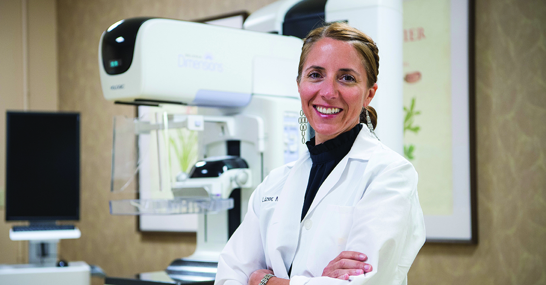
By Dr. Eva Lizer, Women's Life Imaging
Radiologists like myself worried about tomosynthesis, also known as 3D mammography, when it first emerged as a tool to look for breast cancer several years ago. We thought it might be more uncomfortable to women, and worried that the amount of radiation used would be too high. We also wondered whether tomosynthesis would be less useful than standard mammography in identifying certain signs of breast cancer. However, three years into our experience with tomosynthesis at Women’s Life Imaging Center, I can’t imagine ever going back to 2D mammography again. Tomosynthesis is really that much better.
Tomosynthesis is an X-ray of the breast, just like a regular 2D mammogram. From the patient’s perspective, the exam itself is nearly identical except it’s just a few seconds longer. Instead of taking one picture at a time, the machine moves through a short arc and takes multiple low-dose X-rays of the breast. A computer puts these together as a 3D set of images. In a 2D mammogram, the radiologist looks at the complex architecture of breast tissue in one flat image. Overlapping normal breast tissue can appear abnormal on a 2D mammogram, but with a 3D mammogram a radiologist can more confidently see that there is no true abnormality. This prevents “callbacks” for additional mammograms or ultrasound. In fact, multiple scientific studies have shown significant reductions in the percentage of women asked to return for more testing – somewhere between 15 and 40 percent. In addition, these studies have found higher cancer detection rates with tomosynthesis. That’s because a 3D mammogram provides better visibility of small cancers that could be hiding in breast tissue.
In the past, a 3D mammogram did use slightly more radiation than a standard 2D mammogram. However, Women’s Life Imaging now uses the newest type of 3D mammogram available and the radiation dose is essentially the same as for a regular 2D digital mammogram. It’s important to note that for any mammogram, the radiation used is significantly less than the amount of background radiation we are exposed to each year from the environment.
Every year, more than 200,000 women in the United States are diagnosed with breast cancer. According to Centers for Disease Control and Prevention 2013 statistics, New Hampshire has the highest incidence of breast cancer for any state. When breast cancers are found early—before they have spread to other parts of the body—women have a much better chance of surviving. Mammograms are still the best way to screen women for early breast cancers, and I strongly believe that 3D mammography addresses many of the shortcomings of standard mammography. Because there is no better proven way to detect early breast cancers, we recommend annual mammograms beginning at age 40.
Dr. Eva Lizer is a fellowship trained breast imager at Women’s Life Imaging Center, a joint venture between Wentworth-Douglass Hospital and Frisbie Memorial Hospital to provide breast imaging and bone densitometry services. Women’s Life Imaging Center, located in Somersworth, NH, is recognized as a Breast Imaging Center of Excellence by the American College of Radiology.
For more information, visit womenslifeimaging.com or call (603) 742-6673.